Many people start the morning with a cup of coffee to compensate for the lack of sleep, keeping them alert during the day. However, consumption of caffeine may mitigate some of the adverse effects of sleep deprivation as it does not replace a healthy, full night’s sleep.1
Experts in sleep medicine recommend that adults should obtain at least seven hours of sleep per night for optimal health.2 However, the average sleep duration has decreased since 1985, with the percentage of adults sleeping ≤ 6 h increasing by 31%.3 Many people might wonder why sleep has evolved if it prevented feeding and procreation and could expose animals to predators, seemingly contracting principles of the successful survival of a species.4 While the answer remains elusive, some theories propose the necessity of sufficient sleep for optimal functioning. A recent compelling theory called the “brain elasticity theory” suggests that sleep is correlated with changes in the structure and organization of the brain, indicating the essential role of sleep in cognition and memory.5
The two-process model of sleep
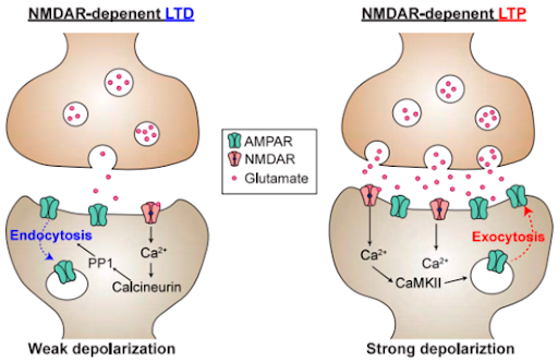
Sleep is an active state of unconsciousness when the brain and body decrease responsiveness to external stimuli.6 Brain plasticity, or neuroplasticity, is the ability of the nervous system to respond to stimuli by reorganizing its structure, functions, or neural connections.7 The synapse refers to the structure that allows one neuron to pass information to another neuron, mediating communication between neurons. Synaptic strength indicates the level of activity of the receiving neuron, or the postsynaptic neuron, in response to the presynaptic neuron. Long-term synaptic plasticity is an activity-dependent change in the transmission of information, modifying synaptic strength by enhancing through long-term potentiation (LTP) or depressing through long-term depression (LTD) (Figure 1).7 Much research has been done on this topic in order to investigate its involvement in various neurological phenomena, such as learning, working memory, brain development, and other neural functions.
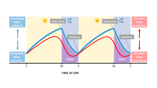
Sleep is regulated by a two-process model consisting of the homeostatic process (S) and the circadian process (C).8 This two-process model of sleep-wake regulation determines the timing of sleep onset and offset in which sleep is triggered when Process S reaches a threshold9 (Figure 2). Process S monitors time awake as sleep pressure builds up during wakefulness and decreases during sleep. Process C is mediated by circadian rhythms, which are internal processes that respond to the environment and regulate sleep-wake cycles. Circadian rhythms repeat roughly every 24 hours in humans,9 representing the daily oscillatory modulation of the threshold levels. The circadian drive for wakefulness increases during the day and declines in the night.
Sleep deprivation (SD) perturbs this natural two-stage process. Neuroimaging studies reveal changes in brain activity, reporting significant reductions in metabolism throughout the thalamic, parietal, and prefrontal regions of the brain, which are involved in cognition and memory.8 SD disrupts normal sleep and negatively affects various aspects of body functioning, and one of the prominent changes of SD is its effect on brain functioning, especially cognition and memory.
The influence of Sleep Deprivation on memory
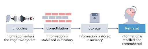
Before understanding the influence of sleep deprivation on memory, it is vital to introduce the concept of memory. There are four stages of memory: encoding, consolidation, storage, and retrieval (Figure 3). Studies have shown how SD affects each stage of memory.
The most well-established neural correlation with insufficient sleep is the hippocampus, a brain structure that plays an essential role in learning and memory.10,11 The hippocampal-dependent memory system is responsible for episodic, or more personal, memories, such as one’s birthday, as opposed to semantic, or fact-based, memories, such as the speed of light. Research has shown that CA1 neurons and the dentate gyrus (DG) in the hippocampus play a critical role in this memory system (Figure 4).12
DG is the input region of the hippocampus, which filters information and has been reported to play a key role in learning, memory, and spatial coding. SD changes the structure of DG neurons by reducing the spine density of DG neurons, which could lead to deficits in memory functions.13
CA1 is the major output region of the hippocampus, and its neurons are involved in the encoding and consolidation of these episodic memories. In 2016, a group of scientists found that SD in mice suppressed the activation of mTORC1, a regulator of cell growth and metabolism, and protein synthesis in hippocampal neurons. Restoring protein synthesis by reactivation of this gene protected mice from memory impairment caused by SD. Moreover, another group of scientists found that depriving mice of sleep for five hours dramatically reduced the connectivity between neurons in the hippocampus, affecting CA1 neurons. Limited sleep caused reduced connectivity between neurons to change the structure and activity of the brain, influencing neuroplasticity. Recovery sleep led to the rewiring of neurons in the hippocampus and restored memory impairments.14 Other studies have also shown that sleep modulates NMDA receptor-dependent signaling in the hippocampus, which is essential for memory and learning. When sleep is limited, this pathway is disrupted, affecting the presentation of NMDA receptors in the synapse that are critical for triggering long-term synaptic plasticity such as LTP or LTD, leading to memory impairment.
Additional research investigating the effect of SD on the frontal and parietal cortices of the brain, both of which are important for cognitive and memory functions, concluded that SD affects working memory—the temporary memory system used to hold a small amount of information for cognitive tasks.15,16 Examples of working memory include remembering a phone number or recalling directions.
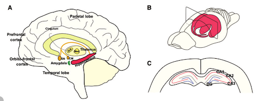
Figure 4. Brain anatomy. (A) Principal anatomy of the human hippocampal memory systems and the brain regions involved in learning and memory. (B) Schematic rat brain with the hippocampal formation highlighted. (C) Schematic hippocampal slice.
How does SD influence the retrieval of memory?
While it is well established that SD after learning impairs hippocampal memory processes, it is unknown whether SD leads to memory loss or suboptimal storage of information that cannot be accessed and retrieved. As a consequence, there is debate on the stage of memory—encoding, consolidation, storage, and retrieval—that SD disrupts. A recent study published in 2023 reported that amnesia is not caused by information loss but is rather a problem of retrievability and can be restored by drug treatment or external stimulation of engram cells, which are the cells activated when a memory is recalled.17
Memory engrams are the physical and chemical changes elicited by learning that underlie newly formed memories. Previous research has shown that neurons activated by an experience become engram cells that later become necessary for successful memory retrieval and expression.18 In fact, memory can be artificially induced by stimulating the engram cells in the brain that encode the experience. For example, studies have reported that memory can be successfully recalled upon stimulation of the memory engram in amnesia patients. 17, 18
In the 2023 study, researchers showed that the hippocampal object-location memories were impaired in sleep-deprived mice using a behavioral paradigm called the object-location memory (OLM) task. This task is based on the innate preference of rodents to spend more time exploring novel objects compared to familiar objects and recognize when an object has been relocated. 19 After training, animals that remember the original training will explore the displaced object relative to the non-displaced object during the testing phase, and spatial memory impairment is measured by the difference in exploration times.
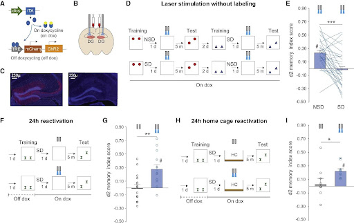
Figure 5 shows the paradigm of the OLM task in the 2023 study. Results show that, while non-SD mice preferred the relocated object, mice subjected to 6 hours of SD explored both objects—the one placed in the original position and the one relocated—to a similar extent, suggesting that they failed to detect the relocation of an object and indicating memory retrieval impairment but not deficits in memory loss. When the researchers activated the DG, the input region of the hippocampus, memory engram responsible for spatial novelty, it became accessible and could be successfully retrieved.17
As most previous studies stated that SD mainly affects the encoding and consolidation stages of memory, sleep loss can potentially perturb memory at the retrieval phase as sleep is known to affect the parietal lobe, which has been shown to contribute to episodic memory retrieval.20 This study, along with other recent studies, has demonstrated that the retrieval stage of memory is impaired after SD, potentially causing memory and cognitive impairments when sleep is hindered.
Conclusion
Sleep deprivation is common in modern society, affecting people of all ages. Although many people have experienced impaired body functioning, including deficits in memory and cognitive functions, few people understand the science behind the lack of sleep. Sleep itself is a complicated topic and involves multiple proposed mechanisms and theories. It is well known in the scientific community that hippocampal-dependent memory is mainly affected by SD, but there is debate on which stage of memory is influenced by SD. While research shows that memory encoding and consolidation were affected, recent studies have revealed the impact of SD on memory retrieval through the representation of memory engrams in the brain. This area of research is ongoing, but it is clear that SD imposes multiple adverse effects on our bodies, significantly influencing brain functioning. It is vital for people to acknowledge the importance of sleep and try to obtain seven hours of sleep every night for optimal health.
References:
- Stepan, M. E., Altmann, E. M., Fenn, K. M., & Link to external site, this link will open in a new window. (2021). Caffeine selectively mitigates cognitive deficits caused by sleep deprivation. Journal of Experimental Psychology: Learning, Memory, and Cognition, 47(9), 1371–1382. https://doi.org/10.1037/xlm0001023
- Watson, N. F., Badr, M. S., Belenky, G., Bliwise, D. L., Buxton, O. M., Buysse, D., Dinges, D. F., Gangwisch, J., Grandner, M. A., Kushida, C., Malhotra, R. K., Martin, J. L., Patel, S. R., Quan, S. F., & Tasali, E. (2015). Recommended Amount of Sleep for a Healthy Adult: A Joint Consensus Statement of the American Academy of Sleep Medicine and Sleep Research Society. Sleep, 38(6), 843–844. https://doi.org/10.5665/sleep.4716
- Ford, E. S., Cunningham, T. J., & Croft, J. B. (2015). Trends in Self-Reported Sleep Duration among US Adults from 1985 to 2012. Sleep, 38(5), 829–832. https://doi.org/10.5665/sleep.4684
- Why Did Sleep Evolve? (n.d.). Scientific American. https://doi.org/10.1038/scientificamericanmind0113-70b
- Mateos-Aparicio, P., & Rodríguez-Moreno, A. (2019). The Impact of Studying Brain Plasticity. Frontiers in Cellular Neuroscience, 13, 66. https://doi.org/10.3389/fncel.2019.00066
- Brinkman, J. E., Reddy, V., & Sharma, S. (2022). Physiology of Sleep. In StatPearls. StatPearls Publishing. http://www.ncbi.nlm.nih.gov/books/NBK482512/
- Alkadhi, K., Zagaar, M., Alhaider, I., Salim, S., & Aleisa, A. (2013). Neurobiological Consequences of Sleep Deprivation. Current Neuropharmacology, 11(3), 231–249. https://doi.org/10.2174/1570159X11311030001
- Achermann P. (2004). The two-process model of sleep regulation revisited. Aviation, space, and environmental medicine, 75(3 Suppl), A37–A43.
- Goel, N., Basner, M., Rao, H., & Dinges, D. F. (2013). Circadian Rhythms, Sleep Deprivation, and Human Performance. Progress in Molecular Biology and Translational Science, 119, 155–190. https://doi.org/10.1016/B978-0-12-396971-2.00007-5
- National Institute of General Medical Sciences. (n.d.). National Institute of General Medical Sciences (NIGMS). Retrieved March 19, 2023, from https://nigms.nih.gov/
- Havekes, R., & Abel, T. (2017). The tired hippocampus: The molecular impact of sleep deprivation on hippocampal function. Current Opinion in Neurobiology, 44, 13–19. https://doi.org/10.1016/j.conb.2017.02.005
- Prince, T.-M., Wimmer, M., Choi, J., Havekes, R., Aton, S., & Abel, T. (2014). Sleep deprivation during a specific 3-hour time window post-training impairs hippocampal synaptic plasticity and memory. Neurobiology of Learning and Memory, 109, 122–130. https://doi.org/10.1016/j.nlm.2013.11.021
- Raven, F., Meerlo, P., Van der Zee, E. A., Abel, T., & Havekes, R. (2019). A brief period of sleep deprivation causes spine loss in the dentate gyrus of mice. Neurobiology of Learning and Memory, 160, 83–90. https://doi.org/10.1016/j.nlm.2018.03.018
- Havekes, R., Park, A. J., Tudor, J. C., Luczak, V. G., Hansen, R. T., Ferri, S. L., Bruinenberg, V. M., Poplawski, S. G., Day, J. P., Aton, S. J., Radwańska, K., Meerlo, P., Houslay, M. D., Baillie, G. S., & Abel, T. (2016). Sleep deprivation causes memory deficits by negatively impacting neuronal connectivity in hippocampal area CA1. ELife, 5, e13424. https://doi.org/10.7554/eLife.13424
- Frenda, S. J., & Fenn, K. M. (2016). Sleep Less, Think Worse: The Effect of Sleep Deprivation on Working Memory. Journal of Applied Research in Memory and Cognition, 5(4), 463–469. https://doi.org/10.1016/j.jarmac.2016.10.001
- Krause, A. J., Simon, E. B., Mander, B. A., Greer, S. M., Saletin, J. M., Goldstein-Piekarski, A. N., & Walker, M. P. (2017). The sleep-deprived human brain. Nature Reviews Neuroscience, 18(7), Article 7. https://doi.org/10.1038/nrn.2017.55
- Bolsius, Y. G., Heckman, P. R. A., Paraciani, C., Wilhelm, S., Raven, F., Meijer, E. L., Kas, M. J. H., Ramirez, S., Meerlo, P., & Havekes, R. (2023). Recovering object-location memories after sleep deprivation-induced amnesia. Current Biology, 33(2), 298-308.e5. https://doi.org/10.1016/j.cub.2022.12.006
- Josselyn, S. A., & Tonegawa, S. (2020). Memory engrams: Recalling the past and imagining the future. Science (New York, N.Y.), 367(6473), eaaw4325. https://doi.org/10.1126/science.aaw4325
- Vogel-Ciernia, A., & Wood, M. A. (2014). Examining Object Location and Object Recognition Memory in Mice. Current Protocols in Neuroscience / Editorial Board, Jacqueline N. Crawley … [et Al.], 69, 8.31.1-8.31.17. https://doi.org/10.1002/0471142301.ns0831s69
- Wagner, A. D., Shannon, B. J., Kahn, I., & Buckner, R. L. (2005). Parietal lobe contributions to episodic memory retrieval. Trends in Cognitive Sciences, 9(9), 445–453. https://doi.org/10.1016/j.tics.2005.07.001
Figures references:
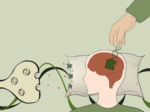
- Cover image: created by author
- Figure 1: Hyun, J. S., Inoue, T., & Hayashi-Takagi, A. (2020). Multi-Scale Understanding of NMDA Receptor Function in Schizophrenia. Biomolecules, 10(8), Article 8. https://doi.org/10.3390/biom10081172
- Figure 2: What makes a good night’s sleep? (n.d.). The Physiological Society. Retrieved March 21, 2023, from https://www.physoc.org/magazine-articles/what-makes-a-good-nights-sleep/
- Figure 3: Roediger, H. L., & Abel, M. (2022). The double-edged sword of memory retrieval. Nature Reviews Psychology, 1(12), Article 12. https://doi.org/10.1038/s44159-022-00115-2
- Figure 4: Temido-Ferreira, M., Coelho, J., Pousinha, P., & Lopes, L. (2019). Novel Players in the Aging Synapse: Impact on Cognition. Journal of Caffeine and Adenosine Research, 9. https://doi.org/10.1089/caff.2019.0013
- Figure 5: Bolsius, Y. G., Heckman, P. R. A., Paraciani, C., Wilhelm, S., Raven, F., Meijer, E. L., Kas, M. J. H., Ramirez, S., Meerlo, P., & Havekes, R. (2023). Recovering object-location memories after sleep deprivation-induced amnesia. Current Biology, 33(2), 298-308.e5. https://doi.org/10.1016/j.cub.2022.12.006